
FIGURE
Fig. 6
Fig. 6
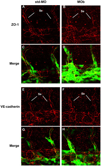
Immunofluorescence analysis by confocal microscopy of endothelial intercellular junctions. Immunofluorescence experiments were performed on tg(fli1:EGFP)y1 embryos at 2 dpf. Confocal images of tail vessels of std-MO (A, E) and MOb injected embryos (B, F) labeled with an anti-ZO-1 and VE-cadherin antibody (red) and the respective merged images (C, D, G, H). Se: intersegmental vessels. |
Expression Data
| Antibodies: | |
|---|---|
| Fish: | |
| Knockdown Reagent: | |
| Anatomical Terms: | |
| Stage: | Long-pec |
Expression Detail
Antibody Labeling
Phenotype Data
Phenotype Detail
Acknowledgments
This image is the copyrighted work of the attributed author or publisher, and
ZFIN has permission only to display this image to its users.
Additional permissions should be obtained from the applicable author or publisher of the image.
Full text @ PLoS One