
Fig. 7
- ID
- ZDB-FIG-130117-39
- Publication
- Sousa et al., 2012 - A new zebrafish bone crush injury model
- Other Figures
- All Figure Page
- Back to All Figure Page
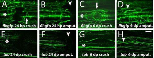
Blood vessels structure was compromised, whereas nerve bundles were not after crush injury. (A–D) Imaging of the transgenic line fli:EGFP that labels the endothelial cells at (A) 24hpc, arrow indicates the main bony ray artery (B) 24hpa (C) 6dpc, arrow highlights the blood vessels mispatterning (D) 6dpa. (E–H) Immunohistochemistry with the antibody anti-acetylated Tubulin to detect nerve fibres at (E) 24hpc (F) 24hpa (G) 6dpc (H) 6dpa. Arrowheads indicate the amputation plane and asterisks indicate crush injury sites. Scale bar corresponds to 100μm in all panels. (hpa – hours post-amputation; hpc – hours post-crush injury; dpa – days post-amputation; dpc – days post-crush injury). |