
FIGURE
Fig. 5
- ID
- ZDB-FIG-130117-37
- Publication
- Sousa et al., 2012 - A new zebrafish bone crush injury model
- Other Figures
- All Figure Page
- Back to All Figure Page
Fig. 5
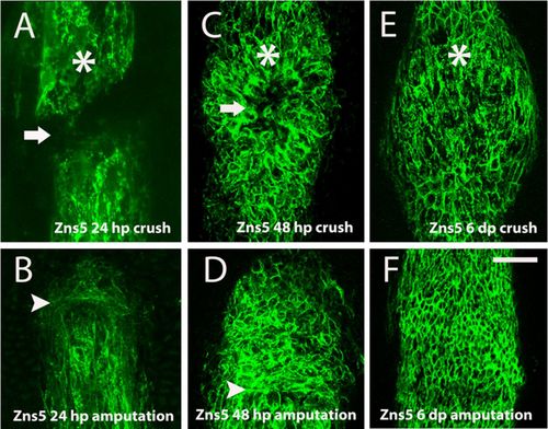
Skeletal cell deposition was delayed and patterning was affected after crush injury. (A–F) Immunohistochemistry with the antibody anti-Zns5 to detect skeletal cells. (A) 24hpc, the arrow highlights the lack of bone cells deposition (B) 24hpa (C) 48dpc (D) 48hpa (E) 6dpc (F) 6dpa, near the amputation plane. Arrowheads indicate the amputation plane and asterisks indicate crush injury sites. Scale bar corresponds to 50µm in all panels. (hpa – hours post-amputation; hpc – hours post-crush injury). |
Expression Data
Expression Detail
Antibody Labeling
Phenotype Data
Phenotype Detail
Acknowledgments
This image is the copyrighted work of the attributed author or publisher, and
ZFIN has permission only to display this image to its users.
Additional permissions should be obtained from the applicable author or publisher of the image.
Full text @ Biol. Open