FIGURE
Fig. 3
- ID
- ZDB-FIG-130110-17
- Publication
- Fieramonti et al., 2012 - Time-gated optical projection tomography allows visualization of adult zebrafish internal structures
- Other Figures
- All Figure Page
- Back to All Figure Page
Fig. 3
|
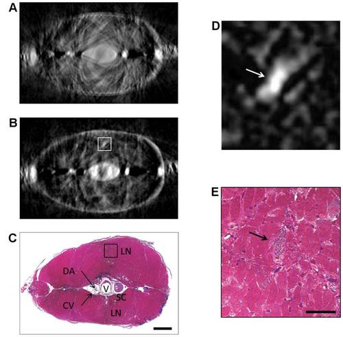
Comparison between transverse sections. (a) Virtual transverse section reconstructed by OPT. (b) Virtual transverse section reconstructed by early-TGOPT. (c) Correspondent histological section for comparison. V: vertebra, SC: spinal cord, LN: lateral nerve, DA: dorsal aorta, CV: cardinal vein. (d) Closeup of the highlighted area in the early-TGOPT section. (e) Closeup of the highlighted area in the histological section. The arrows point to the lateral nerve. Scale bar for (a)-(c) is 400 μm; scale bar for (d)-(e) is 50 μm. |
Expression Data
Expression Detail
Antibody Labeling
Phenotype Data
Phenotype Detail
Acknowledgments
This image is the copyrighted work of the attributed author or publisher, and
ZFIN has permission only to display this image to its users.
Additional permissions should be obtained from the applicable author or publisher of the image.
Full text @ PLoS One

