
Fig. S5
- ID
- ZDB-FIG-121217-36
- Publication
- Nguyen-Chi et al., 2012 - Morphogenesis and Cell Fate Determination within the Adaxial Cell Equivalence Group of the Zebrafish Myotome
- Other Figures
- All Figure Page
- Back to All Figure Page
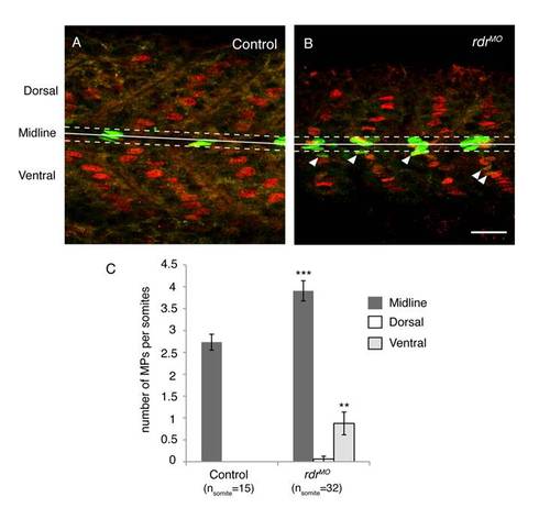
Supernumerary MPs in rdr morphans are mainly localised in the midline and in the ventral region of the myotome. MPs were stained with anti-Eng (green) and prox1 (red) antibodies in uninjected controls (A) and rdr MO injected (B) embryos at 1 dpf. The midline (solid line), midline region (dashed lines), ventral and dorsal regions are shown. Here the midline region corresponds to the 5 µm regions flanking either side of the midline. Scale bar = 25 μm (C) Graphic representation of the number of MPs in the midline, dorsal and ventral regions of the somite in uninjected controls and rdr morphans. ***p<0.001 and **p<0.005, values = means and error bars = S.E.M. |