
FIGURE
Fig. 4
- ID
- ZDB-FIG-121130-44
- Publication
- Issa et al., 2012 - Spinocerebellar ataxia type 13 mutation associated with disease onset in infancy disrupts axonal pathfinding during neuronal development
- Other Figures
- All Figure Page
- Back to All Figure Page
Fig. 4
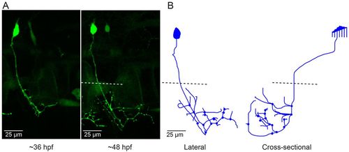
CaP neurons that express aoR3H tend to branch excessively at the distal end of the axon. (A) Projected confocal images obtained from an aoR3H-expressing CaP neuron at 36 and ~48 hpf are shown. At ~48 hpf, the distal axonal arbor is highly branched. (B) Projected Neurolucida trace of the ~48 hpf image stack is shown from lateral (left) and cross-sectional (right) perspectives. See Fig. 1 legend for color code, etc. |
Expression Data
Expression Detail
Antibody Labeling
Phenotype Data
Phenotype Detail
Acknowledgments
This image is the copyrighted work of the attributed author or publisher, and
ZFIN has permission only to display this image to its users.
Additional permissions should be obtained from the applicable author or publisher of the image.
Full text @ Dis. Model. Mech.