
- Title
-
Spinocerebellar ataxia type 13 mutation associated with disease onset in infancy disrupts axonal pathfinding during neuronal development
- Authors
- Issa, F.A., Mock, A.F., Sagasti, A., and Papazian, D.M.
- Source
- Full text @ Dis. Model. Mech.
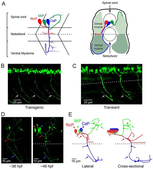
CaP morphology is extremely reproducible. (A) The schematics illustrate the stereotypical morphology and axonal trajectories of the RoP (red), MiP (green) and CaP (blue) primary motor neurons at ~48 hpf from lateral (left) –and cross-sectional (right) perspectives (Myers et al., 1986). Dashed horizontal lines correspond to the horizontal myoseptum (choice point). Left: chevrons show the hemisegment boundaries. Anterior is to the left and dorsal is up. Right: Dorsal is up. (B) Three adjacent hemisegments from a germline transgenic animal [Tg(Mmu.MNX1-MNE:EGFP)] that expresses EGFP in motor neurons were imaged at ~48 hpf. Arrows indicate the characteristic loop of the CaP axon under and around the ventral muscle. In this and subsequent figures, confocal images were projected and lateral or quasi-lateral views are shown. Anterior is to the left and dorsal is up. (C) Two adjacent hemisegments from an animal that was injected with the EGFP plasmid for transient expression in motor neurons were imaged at ~48 hpf. Arrows indicate the characteristic loop of the CaP axon under and around the ventral muscle. In this and subsequent figures, the dashed line indicates the location of the horizontal myoseptum (choice point). (D) Projected confocal images were acquired from an animal transiently expressing EGFP in all three primary motor neurons in one hemisegment at ~36 hpf (left) and ~48 hpf (right). (E) Using the confocal image stack acquired at ~48 hpf, each neuron was traced using Neurolucida. Projected traces are shown from lateral (left) and cross-sectional (right) perspectives. In D and E, and in subsequent figures unless otherwise noted, arrowheads indicate cell bodies and arrows indicate axons: red, RoP; green, MiP; blue, CaP. In E and in subsequent Neurolucida traces, circles indicate branch points. EXPRESSION / LABELING:
|
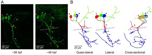
CaP neurons that express exogenous wild-type Kv3.3 develop normally. (A) Shown are projected confocal images of a hemisegment in which all three primary motor neurons express the wild-type Kv3.3-EGFP fusion protein: left, ~36 hpf; right, ~48 hpf. The images are offset from true lateral views. (B) Projected Neurolucida traces obtained from the image stack acquired at ~48 hpf are shown from the original quasi-lateral (left), corrected lateral (center) and cross-sectional (right) perspectives. See Fig. 1 legend for color code, etc. |
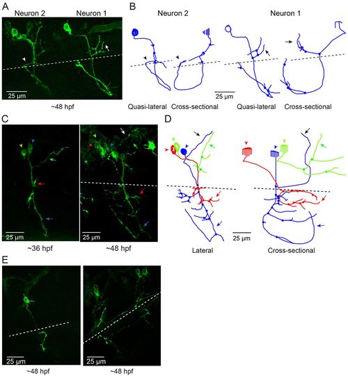
Infant-onset FL mutation causes axonal pathfinding errors in CaP neurons. (A) Projected quasi-lateral image acquired at ~48 hpf shows ioFL-expressing CaP neurons with different axonal abnormalities in adjacent hemisegments. In the right-hand hemisegment, neuron 1 has extended an aberrant collateral into the dorsal myotome (arrow). In the left-hand hemisegment, the main axon shaft of neuron 2 has wrapped around the ventral muscle and then grown into abnormal territory in the dorsal region of the ventral muscle (arrowhead). Note that the other primary motor neurons in the left-hand hemisegment also expressed the ioFL fusion protein. (B) Projected Neurolucida traces obtained from neurons 1 and 2 are shown from quasi-lateral (left) and cross-sectional (right) perspectives. Arrowheads and arrows have the same meaning as in A. (C) Shown are projected confocal images of a hemisegment in which all three primary motor neurons expressed the ioFL-EGFP fusion protein by ~48 hpf: left, ~36 hpf; right, ~48 hpf. Right: the white arrow indicates an aberrant collateral extending from the CaP axon into MiP territory. (D) Projected Neurolucida traces obtained from the ~48 hpf image in C are shown from lateral (left) and cross-sectional (right) perspectives. The black arrow indicates aberrant collateral extending from the CaP axon into MiP territory. (E) Projected confocal images acquired at ~48 hpf show examples of ioFL-expressing neurons with an abnormally truncated axon (left) or axons that could not be traced by Neurolucida (right). See Fig. 1 legend for color code, etc. |
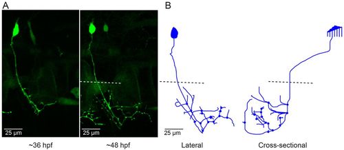
CaP neurons that express aoR3H tend to branch excessively at the distal end of the axon. (A) Projected confocal images obtained from an aoR3H-expressing CaP neuron at 36 and ~48 hpf are shown. At ~48 hpf, the distal axonal arbor is highly branched. (B) Projected Neurolucida trace of the ~48 hpf image stack is shown from lateral (left) and cross-sectional (right) perspectives. See Fig. 1 legend for color code, etc. |