
Fig. 7
- ID
- ZDB-FIG-121025-46
- Publication
- Ben Shoham et al., 2012 - S1P1 inhibits sprouting angiogenesis during vascular development
- Other Figures
- All Figure Page
- Back to All Figure Page
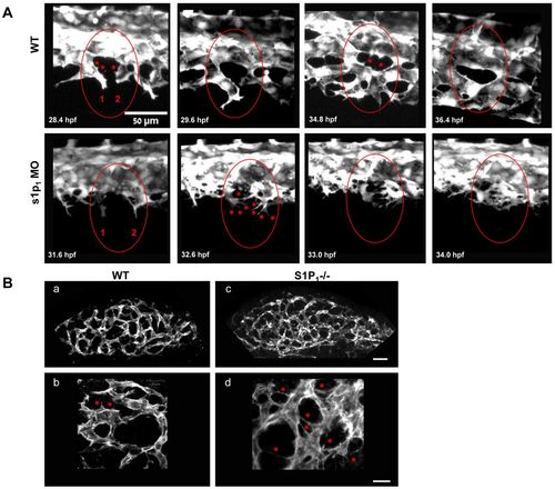
Excessive filopodia formation leads to aberrant vascular remodeling in s1p1 MO zebrafish and S1P1-/- mouse embryos. (A) Sequential confocal microscopy images showing lateral views of the CVPs from WT and s1p1 MO Tg(fli-egfp)y1 zebrafish embryos. Circles demarcate two sprouts, marked as 1 and 2, in WT and s1p1 MO embryos that extend filopodia (marked with asterisks). This results in the formation of a capillary loop in the WT and in over-sprouting and fusion in the mutant. For visualization of the entire sequence, see supplementary material Movies 1 and 2. (B) Immunofluorescent staining with anti-CD31 of whole-mount forelimbs from E9.5 WT (a,b) and S1P1-/- (c,d) mice; b and d show magnifications of a and c, respectively. Asterisks indicate excessive filopodia in S1P1-/- limbs, compared with WT limbs. Scale bars: in a,c, 50 μm; in b,d, 20 μm. |
| Fish: | |
|---|---|
| Knockdown Reagent: | |
| Observed In: | |
| Stage: | Prim-15 |