|
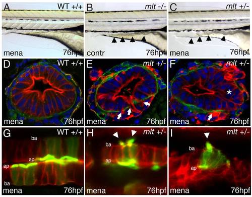
Oxidative stress induces invasive remodeling in mlt heterozygous larvae. (A–C) Lateral images of live WT (A), mlt homozygous (B), and mlt heterozygous larvae (C). The WT and mlt heterozygous larvae received 3 h of treatment with Menadione beginning at 73 hpf. Menadione treated heterozygote (C) larvae have an intestinal phenotype (arrowheads) resembling the untreated mlt homozygous larvae (B). (D–F) Corresponding histological cross-sections (representative of larvae in A, C) with intestinal epithelial cells labeled red (anti-keratin immunostain) and basement membrane in green (anti-laminin immunostain). Menadione causes epithelial cell invasion (arrows) and stratification (asterisks) in mlt heterozygous larvae (E, F) but does not affect epithelial architecture in the WT intestines (D). (G–I) Sagittal confocal scans through the intestine of WT and mlt heterozygotes treated with menadione. Both larvae express LifeAct-GFP in a subset of intestinal epithelial cells. Actin-rich invadopodia-like protrusions (green) are seen arising from the basal epithelial cell membrane of menadione treated heterozygous larvae (arrowheads, H, I). Actin is located nearly exclusively in the apical brush border of WT epithelial cells (G): Red -membrane mCherry; ba, basal epithelial cell border; ap, apical epithelial cell border.
|