
Fig. 2
- ID
- ZDB-FIG-120914-19
- Publication
- Gupta et al., 2012 - A Splice Site Mutation in Laminin-alpha2 Results in a Severe Muscular Dystrophy and Growth Abnormalities in Zebrafish
- Other Figures
- All Figure Page
- Back to All Figure Page
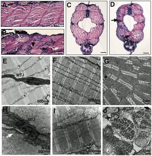
Abnormal myosepta and myofiber detachment in laminin-α2 deficient lama2cl501/cl501 fish. (A & B) Hematoxylin and eosin staining of longitudinal sections of wild-type and lama2 mutant fish at 5 dpf. Mutant muscles showed highly disorganized myofibers in the affected somites with irregular myosepta boundaries (arrowhead) and eosin positive detached myofibers (arrow). (C–D) Cross-sections of wild-type and mutation fish also showed smaller myotome and degenerating muscle fibers in lama2 mutant fish at 5 dpf (arrows), bars = 10 μm. (E & H) Electron microscopy showed myofiber detachment from the myotendinous junction (MTJ) in mutant muscles (arrow). (F & I) The myofibers in wild-type muscles attached tightly to the surrounding fibers while mutant muscle displayed large gaps in the extracellular matrix between adjacent fibers and disorganized Z- lines (black arrow) and M-lines (while arrow). (G & J) Defects in extracellular-matrix results in damaged myofibers in the myotome (cross-section). A large number of apoptotic nuclei were observed in the mutant muscles (J, arrow). |
| Fish: | |
|---|---|
| Observed In: | |
| Stage: | Day 5 |