Fig. 8
- ID
- ZDB-FIG-120831-23
- Publication
- Mu et al., 2012 - Visual Input Modulates Audiomotor Function via Hypothalamic Dopaminergic Neurons through a Cooperative Mechanism
- Other Figures
- All Figure Page
- Back to All Figure Page
|
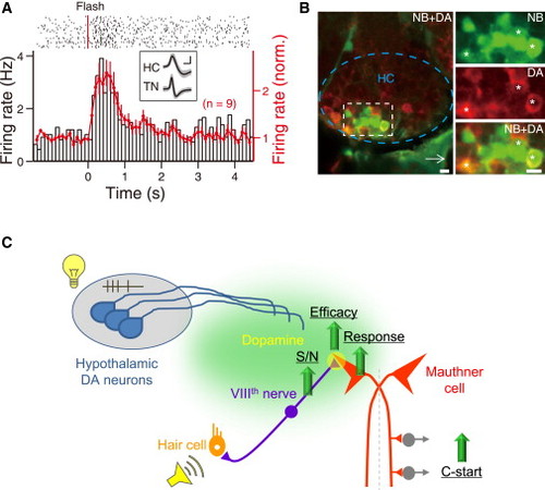
Dopaminergic Neurons in the Caudal Hypothalamus Exhibit Bursting Activity in Response to Flash Stimulus and Send Projections Nearby the VIIIth Nerve-Mauthner Cell Circuit (A) Example (top; left y axis in bottom, black open bars) and summary (right y axis in bottom, red line) of data showing the spiking activity of HC neurons in response to a 15 ms flash (red line, top). Top, raster plot of a sample recording. Bottom: PSTH of the raster plot (left y axis) and summary of data obtained from 9 larvae (right y axis). Inset, sample (gray) and average (black) waveforms of action potentials from a HC neuron (“HC”) and an optic tectum neuron (“TN”). (B) Neurobiotin-based retrograde tracing of HC dopaminergic neurons. The area of the white square in (left) is enlarged in (right). The dashed blue line and white arrow in (left) mark the location of HC and the tip of notochord, respectively. Top right, NB-ir signal (green). Middle right, DA-ir signal (red). Bottom right, merged signals. The stars indicate double positive HC cells. The images were taken from one optic section with a thickness of <4 μm. Scale represents 0.5 mV, 1 ms in the inset of (A), 5 ¼m in (B). (C) Proposed model for the visual modulation of audiomotor function. A preceding flash induces bursting activities in hypothalamic dopaminergic neurons, which may release dopamine in the vicinity of VIIIth nerves-Mauthner cell circuits. Through D1R activation, the S/N ratio of VIIIth nerve spiking activity is elevated by reducing spontaneous activities, and the efficacy of VIIIth nerve-Mauthner cell synapses is increased, leading to an increase in sound-evoked responses of Mauthner cells and enhancement of auditory C-start escape behavior. See also Figure S7. |
| Antibody: | |
|---|---|
| Fish: | |
| Anatomical Terms: | |
| Stage Range: | Day 4 to Day 6 |

