
Fig. S2
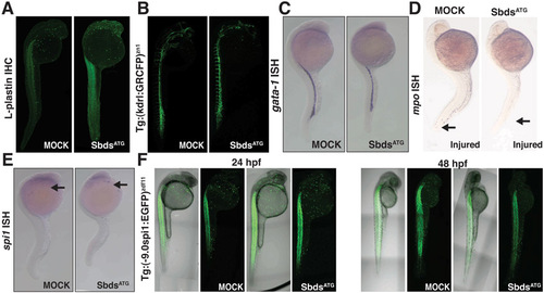
Further characterization of the hematopoietic defects in SbdsATG MO-injected embryos. (A) Immunofluorescence for L-plastin identifies macrophages at 24 hpf in both SbdsATG MO-injected and control embryos. (B) Normal development of vasculature in both SbdsATG MO-injected and control embryos as demonstrated by Tg(kdrl:GRCFP)zn1 transgene. (C) In situ hybridization for gata1, a marker of erythroid progenitors, is identical in control and SbdsATG MO-injected embryos at 24 hpf. (D) Epithelial injury does not recruit neutrophils in SbdsATG MO-injected embryos. Tails were wounded (arrow) at 24 hpf, and neutrophils were visualized at 30 hpf by in situ hybridization for mpo. (E) In situ hybridization for spi1, a marker of common neutrophil/macrophage progenitors, is identical in control and SbdsATG-MO embryos at 24 hpf (arrows). (F) Expression of Tg 9.0 spi1:eGFP at 24 hpf and 48 hpf shows no change in the number or localization of neutrophil/macrophage progenitor markers in SbdsATG MO-injected embryos. Note that expression of eGFP in the muscle is not related to hematopoietic development. |