
Fig. 7
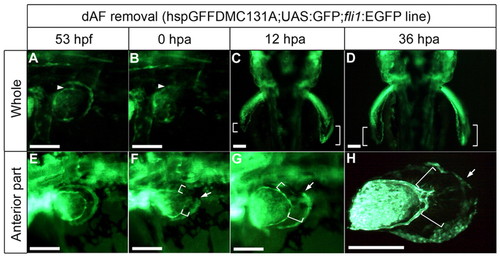
Distal AF removal affected AF outgrowth. The dAF is visualized in the hspGFFDMC131A;UAS:GFP line; the AF base is shown by fli1:EGFP (arrowheads). (A-C) When the dAF was completely removed from the left fin (B), pAF (GFP-negative) development was delayed compared with the control side (C) (n=3/3; white brackets). (D) By 36 hours post-amputation (hpa), the AF length (white brackets) had returned to normal. (E-G) When the anterior part of the dAF was removed (E,F), only the anterior pAF development was delayed (n=6/8; compare with white brackets in E,F). (H) By 36 hpa, the fin shape had returned to normal. Arrows represent the removed/unremoved boundary. The pectoral fin bud was dissected from the fish body and mounted on a glass slide. Scale bars: 200 μm in A,B,E-H; 20 μm in C,D. |