
Fig. 3
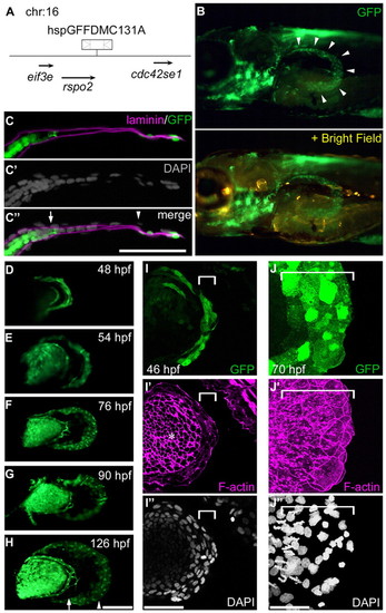
GFP distribution within the distal AF in the hspGFFDMC131A;UAS:GFP line. (A) The hspGFFDMC131A insertion is located 6.6 kb downstream of the R-spondin 2 (rspo2) gene on chromosome 16. (B) An hspGFFDMC131A embryo crossed with the UAS:GFP line. The panel is a lateral view of the pectoral fin bud at 76 hpf; the GFP-positive region is within the edge of the AF (arrowheads). (C-C3) One section of the pectoral fin at 76 hpf (B) processed for three-color immunostaining [C, GFP (green) and Laminin α5 (magenta); C2, DAPI (gray); C3, merged image]. Scale bar: 50 μm. Arrow and arrowhead indicate the fin blood vessel and pAF/dAF boundary, respectively. (D-H) Lateral view of the pectoral fin buds. Scale bar: 100 μm. Arrow indicates the fin blood vessel. Arrowhead indicates the pAF/dAF boundary. (I-J3) High-magnification views of dAF cells (white brackets) at 46 (I-I3) and 70 (J-J3) hpf. Asterisk indicates not the AF but the endoskeletal region. Scale bars: 50 μm. |