|
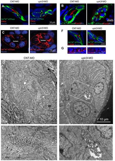
Disruption of epithelial polarity in upk3l morphants. (A–B) Localization of Prkcz (A) or Pard3 (B), Na+/K+-ATPase, and nuclei in cross-sectioned embryos treated with control (CNT-MO) or upk3l MO (upk3l-MO). (C) Distribution of Na+/K+-ATPase and nuclei in 2-dpf control or morphant embryos. (D–E) TEM analysis of the proximal segment of PTs from control or morphant embryos. (D) Cross section of tubule show that it is comprised of 5–6 cells surrounding a central lumen. (E) At higher magnification, control lumens were filled with microvilli (mv) and cilia (ci), whereas microvilli were absent in morphants. (F) Immunolocalization of phosphorylated ERM proteins (P-ERM) in frozen sections of embryos. Sections were co-stained for Na+/K+-ATPase and nuclei. (G) Immunostaining of cilia-associated acetylated-tubulin and nuclei in the PT of laterally oriented embryos. p: proximal tubule, d: distal tubule.
|