
Fig. 2
- ID
- ZDB-FIG-120710-32
- Publication
- Carlin et al., 2012 - Six3 cooperates with Hedgehog signaling to specify ventral telencephalon by promoting early expression of Foxg1a and repressing Wnt signaling
- Other Figures
- All Figure Page
- Back to All Figure Page
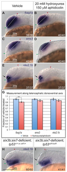
Cellular proliferation and apoptosis do not significantly contribute to reduction of ventral telencephalon. (A-F) Expression of foxg1a (A,B), emx3 (C,D), and nkx2.1b (E,F) at the 20-somite stage in wild-type embryos treated with 2% dimethyl sulfoxide alone (A,C,E) or 20 mM hydroxyurea and 150 μM aphidicolin at 80% epiboly (B,D,F). White bracket indicates length of DV domain measured for quantification. Embryos are shown in lateral view with anterior towards the left. Red and green arrowheads indicate dorsal and ventral edges of the telencephalon, respectively. (G) Graph shows expression domain length along the DV telencephalic axis divided by average DV domain length of vehicle-treated embryos. For each sample, n=11 embryos. Blue and red columns denote vehicle- and hydroxyurea/aphidicolin-treated embryos, respectively. Error bars denote s.e.m. **P<0.01. (H,I) Expression of nkx2.1b at 24 hpf in six3b;six7-deficient embryos (H) that are also tp53zdf1/zdf1 (I). Arrows in E,F,H,I indicate ventral telencephalon. Scale bars: 100 μm. |