
Fig. 6
- ID
- ZDB-FIG-120601-18
- Publication
- Chablais et al., 2012 - The regenerative capacity of the zebrafish heart is dependent on TGFβ signaling
- Other Figures
- All Figure Page
- Back to All Figure Page
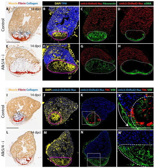
Inhibition of Alk5/4 impairs the production of ECM proteins in the post-infarct. Analyses of heart sections from transgenic fish expressing nuclear DsRed2-Nuc under the cmlc2 cardiomyocyte-specific promoter at 14 dpci. Dashed lines represent the injured areas. DsRed2-Nuc is in red (C,D,G,H) or blue (J-K′,M-N′). (A-D,I-K′) Adjacent sections of control hearts. (E-H,L-N′) Adjacent sections of Alk5/4-i-treated hearts. (A,I) AFOG staining visualizes abundant collagen fibers (blue) at the injury site of control hearts. (E,L) Inhibition of TGFβ/Activin signaling blocks collagen deposition. (B,F) The injury site is demarcated by the absence of Tropomyosin protein (blue). The post-infarct is larger in hearts treated with the drug (F) than in control (B). DAPI (yellow) labels all nuclei. (C,G) Exposure to Alk5/4-i (G) attenuates Fibronectin production (green) in comparison to control (C). (D,H) α-smooth muscle actin-positive cells (αSMA, green) equally surround the post-infarct border in control and drug-treated hearts. (J,M) The injury site is demarcated by the absence of cmlc2::DsRed2-Nuc (blue). DAPI (yellow) labels all nuclei. (K,K′,N,N′) The contra-adhesive matrix protein Tenascin C (TNC, red) is predominantly upregulated at the infarct-myocardium interface in control hearts (K,K′). Treatment with Alk5/4-i completely abolished Tenascin C expression, but increased Vimentin-positive tissue (green; N,N′). (K′,N′) Higher magnifications of the boxed areas in K,N. Arrow (K′) indicates the invasion of new myocardium. Scale bars: 300 μm. |