
Fig. 6
- ID
- ZDB-FIG-120522-41
- Publication
- Liu et al., 2002 - A defect in a novel Nek-family kinase causes cystic kidney disease in the mouse and in zebrafish
- Other Figures
- All Figure Page
- Back to All Figure Page
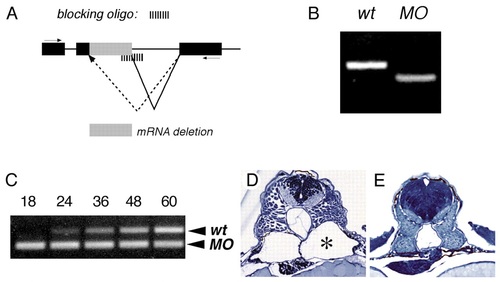
Pronephric cyst formation in Nek8 antisense oligonucleotide injected zebrafish embryos. (A) Splice donor blocking oligo (see Materials and Methods) disruption of Nek8 mRNA splicing. Sequence analysis of the resulting aberrant Nek8 mRNA in 24 hpf embryos by RT-PCR (B) showed that this oligo induced a 129 nucleotides (43 amino acid) in-frame deletion corresponding to the first RCC1 homology domain C-terminal to the Nek kinase domain. (C) Time course quantification of oligo efficacy showed complete absence of wild-type mRNA at 18 hpf and a gradual recovery of wild-type message by 24-60 hpf. (D) Histological analysis of embryos injected as in C show severe pronephric cysts (*) at 60 hpf. (E) Injection of control random oligo (500 nM cytoplasmic concentration) or lower doses of the splice donor blocking oligo (50 nM cytoplasmic concentration) produces normal pronephric kidney formation at 60 hpf. |
| Fish: | |
|---|---|
| Knockdown Reagents: | |
| Observed In: | |
| Stage: | Pec-fin |