
Fig. 2
- ID
- ZDB-FIG-120508-66
- Publication
- Zhang et al., 2012 - Laser ablation of the sonic hedgehog-a-expressing cells during fin regeneration affects ray branching morphogenesis
- Other Figures
- All Figure Page
- Back to All Figure Page
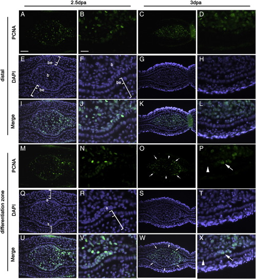
Localized cell proliferation accompanies branch formation. Cell proliferation was detected by immunofluorescence (green) using the proliferating cell nuclear antigen (PCNA) antibody on transverse sections of fin regenerates at 2.5 dpa (A, B, M and N) and 3 dpa (C, D, O and P). DAPI staining (blue) of the same sections is shown in E–H and Q–T. The merged images of the corresponding PCNA and DAPI staining are shown in I–L and U–X. B, F, J, N, R, V, D, H, L, P, T, and X are enlargement of small areas of A, E, I, M, Q, U, C, G, K, O, S, and W, respectively. (A and B) In sections corresponding to the distal part of the fin regenerate at 2.5 dpa, PCNA positive cells are abundant in the blastema. There is no PCNA + cells in the epidermis. (C and D) At 3 dpa, the distribution of PCNA + cells in the distal regenerate is similar to that in (A). (M and N) At 2.5 dpa, in sections corresponding to the differentiation zone, PCNA + cells are more concentrated in the scleroblasts lining the epidermis than in the center of the connective tissue. (O and P) At 3 dpa, as at 2.5 dpa (M), there is a higher concentration of PCNA + cells in scleroblasts than in the central part of the ray but in contrast to 2.5 dpa, the proliferating scleroblasts are localized on the two lateral sides of the hemiray. Note: Bright PCNA staining corresponds to cells that are actively proliferating at the time of tissue fixation. Some residual staining in cells of the connective tissue reflects the past proliferative state of these cells. Arrows in (O, P, W and X) indicate the localized proliferation of the scleroblasts. Arrowheads in (O, P, W and X) indicate the central scleroblasts that show no PCNA signal. Brackets in E, F, Q and R indicate the epidermis. Scale bars: A, E, I, M, Q, U, C, G, K, O, S and W (shown in A) = 20 μm; B, F, J, N, R, V, D, H, L, P, T and X (shown in B) = 7 μm. be: basal layer of epidermis; b: blastema; s: scleroblast. |
Reprinted from Developmental Biology, 365(2), Zhang, J., Jeradi, S., Strähle, U., and Akimenko, M.A., Laser ablation of the sonic hedgehog-a-expressing cells during fin regeneration affects ray branching morphogenesis, 424-433, Copyright (2012) with permission from Elsevier. Full text @ Dev. Biol.