
Fig. 1
- ID
- ZDB-FIG-120508-65
- Publication
- Zhang et al., 2012 - Laser ablation of the sonic hedgehog-a-expressing cells during fin regeneration affects ray branching morphogenesis
- Other Figures
- All Figure Page
- Back to All Figure Page
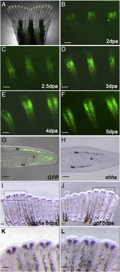
GFP expression recapitulates the endogenous shha expression in the zebrafish transgenic line 2.4shha:gfpABC#15. (A) Bright field and fluorescent images of the caudal fin merge show GFP expression in an intact fin of a 2.4shha:gfpABC#15 transgenic fish. (B–F) Higher magnification of whole-mount images of a few fin rays at different stages of regeneration. (B) At 2 days post amputation (dpa), GFP is expressed in a few cells at the level of the blastema. (C) At 2.5 dpa, GFP expression is restricted to one single domain. (D) At 3 dpa, cleavages start to appear at the proximal end of the GFP-expressing domain and in some rays these cleavages are extending distally. (E) At 4 dpa, a clear separation is visible, and in (F) it is more prominent at 5 dpa. (G) Longitudinal section of a fin regenerate of a 2.4shha:gfpABC#15 transgenic fish at 4 dpa shows GFP expression in the basal layer of the epidermis. (H) In situ hybridization on a longitudinal section of a 4 dpa fin regenerate shows endogenous shha expression in the basal layer of the epidermis. (I–L) In situ hybridization performed on the two lobes of the same fin regenerate at 6 dpa show that shha (I and K) and gfp (J and L) are expressed in the same cell population. The areas indicated by black boxes in I and J are enlarged in K and L. Scale bars in G and H: 20 μm; all other panels: 100 μm. be: basal layer of epidermis; b: blastema. |
Reprinted from Developmental Biology, 365(2), Zhang, J., Jeradi, S., Strähle, U., and Akimenko, M.A., Laser ablation of the sonic hedgehog-a-expressing cells during fin regeneration affects ray branching morphogenesis, 424-433, Copyright (2012) with permission from Elsevier. Full text @ Dev. Biol.