
Fig. S1
- ID
- ZDB-FIG-120427-17
- Publication
- Singh et al., 2012 - Regeneration of amputated zebrafish fin rays from de novo osteoblasts
- Other Figures
- All Figure Page
- Back to All Figure Page
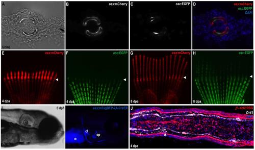
Osteoblast-Specific Transgenic Reporter and Lineage-Labeling Strains (A-D) Transverse section through an uninjured caudal fin from an osx:mCherry; osc:EGFP double transgenic animal. Fin hemirays are outlined by osx:mCherry+ osteoblasts, most of which are also positive for osc:EGFP fluorescence. (E-H) Regenerating fins of osx:mCherry; osc:EGFP animals shown at 4 (E, F) and 8 (G, H) dpa. osx:mCherry is easily visible in the regenerate by 4 dpa, while osc:EGFP fluorescence emerges at 7-8 dpa. Arrowheads indicate amputation planes. (I) Bright field (left) and fluorescent (right) images of a 6 dpf osx:CreER larva, indicating blue mTagBFP fluorescence in bone structures. Asterisk, yolk autofluorescence. cl, clethium; op, opercule. (J) Section through a 4 dpa β-act2:RSG fin regenerate. DsRed fluorescence is detectable in most cells, indicating that the line is suitable for genetic fate-mapping of fin tissues. |
Reprinted from Developmental Cell, 22(4), Singh, S.P., Holdway, J.E., and Poss, K.D., Regeneration of amputated zebrafish fin rays from de novo osteoblasts, 879-886, Copyright (2012) with permission from Elsevier. Full text @ Dev. Cell