Fig. 3
- ID
- ZDB-FIG-120427-14
- Publication
- Singh et al., 2012 - Regeneration of amputated zebrafish fin rays from de novo osteoblasts
- Other Figures
- All Figure Page
- Back to All Figure Page
|
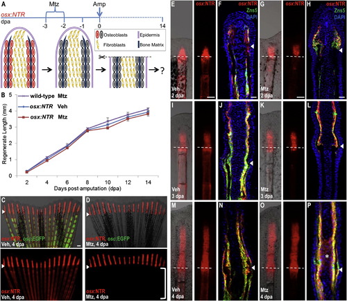
Osteoblast-Depleted Fins Regenerate Normally after Amputation (A) Cartoon summarizing strategy to assess regeneration of amputated fins after genetic ablation of osteoblasts. (B) Lengths of fin regenerates after osteoblast ablation and amputation. As a negative control, wild-type animals were treated with Mtz 2 days before amputation and 4 days after amputation (wild-type, Mtz). osx:NTR animals treated with vehicle (osx:NTR, Veh) or Mtz (osx:NTR, Mtz) prior to amputation regenerated fins with similar efficacy. Data are mean ± SEM from 15 animals each. (C and D) osx:NTR; osc:EGFP animals had indistinguishable regenerative lengths at 4 dpa whether or not osteoblasts were present prior to amputation, and indistinguishable osterix-driven expression in the regenerate. Osteoblast depletion proximal to the amputation plane is evident in Mtz-treated animals by the absence of marker expression (D; bracket). Bottom images show osx:NTR fluorescence only. Arrowheads indicate the plane of amputation. Scale bar = 100 μm. (E–P) Whole-mount views and longitudinal sections of fins at 2 (E–H), 3 (I–L), and 4 (M–P) dpa, highlighting osterix-driven NTR fluorescence. osx:NTR is undetectable below the amputation plane of fins from Mtz-treated animals at 3 and 4 dpa, except for a trail of fluorescent cells at the amputation site. Tissue sections indicate expression of osx:NTR in osteoblasts at each of the 3 time points, and ectopic osx:NTR fluorescence in intraray fibroblasts at 4 dpa in the Mtz treated group (asterisk in P). Dotted lines and arrowheads indicate the plane of amputation. Scale bar = 100 μm. See Figure S3. |
Reprinted from Developmental Cell, 22(4), Singh, S.P., Holdway, J.E., and Poss, K.D., Regeneration of amputated zebrafish fin rays from de novo osteoblasts, 879-886, Copyright (2012) with permission from Elsevier. Full text @ Dev. Cell

