
Fig. 6
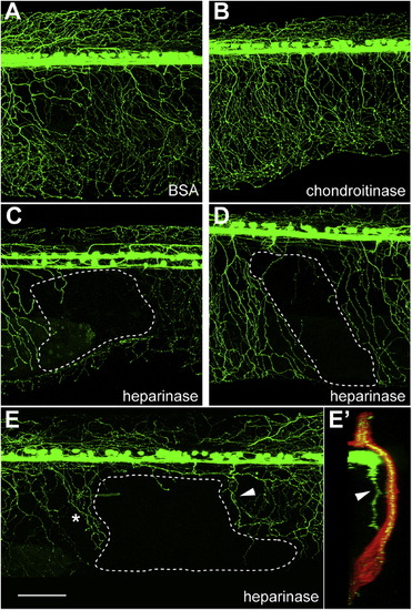
Peripheral Axons Avoid Heparinase III-Injected Areas(A–E) Lateral views of 2 dpf embryos injected with BSA (A) or chondroitinase ABC (B). These embryos did not exhibit obvious defects in RB neuron projections. However, RB neuron peripheral axons often did not innervate heparinase III-injected areas (C–E), suggesting that peripheral axons prefer regions with higher levels of heparan sulfate.(E2) A 90° rotation of (E). Arrowheads in (E) and (E2) indicate an axon that innervated internal tissue. * indicates another axon with a branches beneath the skin. White dotted lines indicate under-innervated areas. Scale bar represented 100 μm. See also Figure S5 and mmc6VIDEO and mmc7VIDEO. |