
Fig. 4
- ID
- ZDB-FIG-120316-70
- Publication
- Brusegan et al., 2012 - ccdc80-l1 Is Involved in Axon Pathfinding of Zebrafish Motoneurons
- Other Figures
- All Figure Page
- Back to All Figure Page
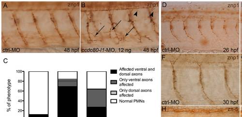
Analysis of motoneurons morphology by means of znp1- and zn-5-immunohistochemistry. (A, B) At 48 hpf, using 12 ng/embryo of morpholino, both ventral (arrows) and dorsal axons (arrowheads) were mis-orientated and over-branched in morphants (B) in comparison to control embryos (A). (C) Statistical analysis showing the percentages of the different phenotypes (affected ventral axons, dorsal axons or both) occurring in control embryos and in morphants, when different doses of ccdc80-l1-MO were injected (12 ng/embryo and 8 ng/embryo). Using a lower dose of morpholino (8 ng/embryo), we observed that in a significant percentage of embryos only ventral axons were defective. (D–G) Immunohistochemistry performed at 26 hpf (D, E) and 30 hpf (F, G) confirmed that loss-of-ccdc80-l1-function affects both CaPs (arrows) and MiPs (arrowheads) axonal migration. (H, I) The same analysis performed at 48 hpf using zn-5 antibody revealed that also SMNs axonal migration is impaired in morphants (arrows in I) in comparison to control embryos (H). (A, B; D–I) Lateral flat-mount preparation was applied for a better visualization of the motoneurons. Lateral views of the trunk region overhanging the yolk extension, dorsal is up and anterior is left. |
| Fish: | |
|---|---|
| Knockdown Reagent: | |
| Observed In: | |
| Stage Range: | Prim-5 to Long-pec |