
Fig. S5
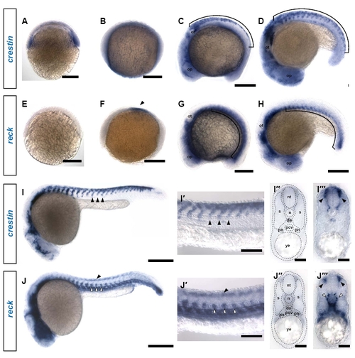
reck is expressed in vasculature and other ventral tissues throughout early development. (A) crestin expression can be detected at shield stage. Scale bar: 200 μm. (B) crestin expression is faint and diffuse at bud stage. Scale bar: 200 μm. (C) crestin is strongly expressed in a dorsal domain corresponding to the premigratory neural crest (bracket) (9 somites). op, optic placode; ot, otic placode. Scale bar: 200 μm. (D) crestin expression is maintained in the neural crest as it begins migrating in dorsoventral streams (bracket) (18 somites) Scale bar: 200 μm. (E) reck is not yet expressed at shield stage. Scale bar: 200 μm. (F) reck expression is initiated at bud stage in a bilateral patch flanking the midline (arrowhead). Scale bar: 200 μm. (G) reck expression expands to the somitic mesoderm (bracket) (9 somites) Scale bar: 200 µm. (H) reck expression remains restricted to the ventral somitic mesoderm (bracket) (18 somites) Scale bar: 200 μm. (I) At 24 hpf, crestin expression is restricted to streams of neural crest migrating ventrally (arrowheads). Scale bar: 250 μm. (I2) High magnification image of the embryo in I showing streams of migrating neural crest (arrowheads). Scale bar: 100 μm. (I22) Section through the trunk of an embryo subjected to crestin in situ hybridization; anatomical references are overlaid. da, dorsal aorta; n, notochord; nt, neural tube; pcv, posterior cardinal vein; pn, pronephros; s, somite; ye, yolk extension. (I222) In this cross-section, streams of neural crest migrating between the somite and the neural tube are defined by crestin expression (arrowheads); these ventromedially migrating cells give rise to DRG. Scale bar: 30 μm. (J) At 24 hpf, reck expression is strongest in the vasculature (white arrowheads), but a faint dorsal expression domain is also apparent (black arrowhead). Scale bar: 250 μm. (J2) High magnification image of the embryo in J showing reck expression in the ventrally situated vasculature (white arrowheads) as well as the fainter dorsal expression domain (black arrowhead). Scale bar: 100 μm. (J22) Section through the trunk of an embryo subjected to reck in situ hybridization; anatomical references are overlaid. (J222) In cross-section, reck expression is evident in both the developing vasculature (white arrowheads) and in a dorsolateral domain consistent with neural crest (black arrowheads). Scale bar: 30 μm. |