|
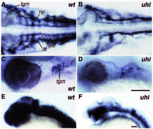
Analysis of neural cell types in mutant uchu hikoushim172 at 30 hpf. (A,C,E) Wild-type. (B,D,F) uchu hikoushim172. (A,B) Dorsal view of znp-1 antibody staining pattern in hindbrain region. Neuropils in each hindbrain segment are affected in mutants. (C,D) Lateral view of Islet-1 antibody staining pattern caudal to the eye. The number of trigeminal ganglion cells is reduced in mutant embryos. (E,F) Lateral view of zrf-1 antibody staining pattern in head region. Mutant embryos have severly reduced amounts of radial glia cells. np, neuropil in hindbrain segment; llf, lateral longitudinal fascicle; mlf, medial longitudinal fascicle; tgm, trigeminal ganglion. Scale bars, (A-D), 100 µm; (E,F) 100 µm.
|