|
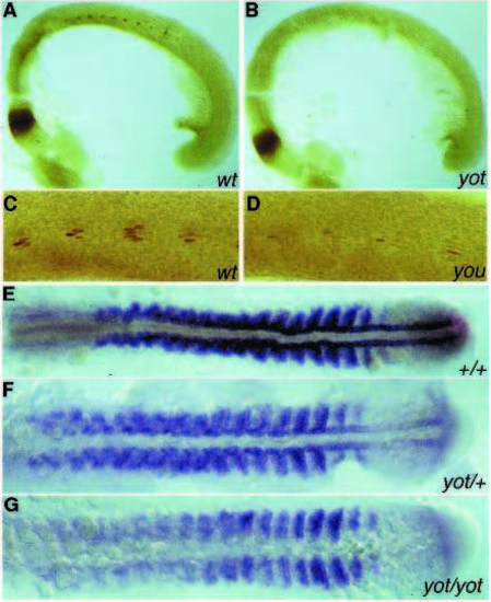
(A) Engrailed (4D9) staining of a 21-somite stage wild-type embryo. Staining is visible at the midbrain-hindbrain boundary and in the somites. In every somite about 2-5 nuclei, representing the muscle pioneers, are stained. In a yot mutant at the same stage (B), staining of the muscle pioneers is absent. (C) 4D9 staining of a wildtype sibling and (D) the weakest you-type mutant, youtm146c. Approximately one Engrailed-positive cell is present per segment. (E-G) myoD-staining of a wild type, a yot mutant and a putative yot/+ embryo at the 17-somite stage, respectively. In the homozygous yot mutant, adaxial staining is absent and the staining in the rest of the paraxial mesoderm is reduced (G). In the yot heterozygous embryo, expression in the adaxial cells is reduced, especially in the presomitic mesoderm, anterior to the tailbud (F).
|