|
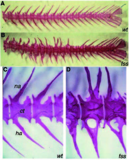
Skeletal phenotype of fss mutants. Skeletal staining on adult wild type (A) and fss mutant (B) tail vertebrae. (C,D) Enlargement of wild-type and mutant vertebrae, respectively at a similar A/P level. ct; centrum, na; neural arch, ha; hemal arch. The centra in fss are almost normal in number and shape, but the length of individual centra is slightly more variable (B). In the wild type (C) every vertebra has four half arches, forming the neural arch on the dorsal side and the hemal arch on the ventral side. In fss mutants (B,D), arches grow out irregularly. For example, on the ventral side of the middle vertebra in D, three half arches are present, two at the left side which fuse, project to the right side, and fuse with the third half arch.
|