
Fig. 3
- ID
- ZDB-FIG-120228-18
- Publication
- Lin et al., 2012 - Quercetin-4'-O-beta-D-glucopyranoside (QODG) Inhibits Angiogenesis by Suppressing VEGFR2-Mediated Signaling in Zebrafish and Endothelial Cells
- Other Figures
- All Figure Page
- Back to All Figure Page
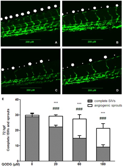
QODG inhibits blood vessel formation in ISVs of zebrafish embryos. (A) Vehicle control: Tg(fli1:EGFP) zebrafish embryos were treated with 0.1% DMSO from 6 hours post fertilization (hpf) to 72 hpf. All intersegmental vessels (ISVs) in the vehicle control group have fully extended to form the dorsal longitudinal anastomotic vessels (DLAVs) at 72 hpf. (B–D) QODG-treated groups: Tg(fli1:EGFP) zebrafish embryos were treated with various concentrations of QODG (20, 60, 180 μM) from 6 hpf to 72 hpf. Pentagons indicate the sites of complete ISVs in zebrafish embryos for all figures, and asterisks indicate the sites of angiogenic sprouts in zebrafish embryos for all figures. (E) Quantitative comparison of blood vessel formation in the vehicle control group and QODG-treated groups. Data are expressed as mean ± SD from three independent experiments. *, the number of complete ISVs in QODG-treated group compared with that in the vehicle control group; ***, P<0.001 vs. vehicle control. #, the number of angiogenic sprouts in QODG-treated group compared with that in the vehicle control group; ###, P<0.001 vs. vehicle control. Scale bars, 200 μm. |