
Fig. 3
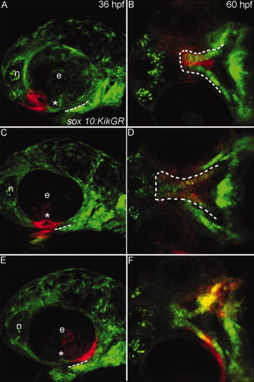
Progenitor domains for the zebrafish palate. A–F: Sox10:KikGR photoconverted cranial neural crest cells (CNCC) in red. A,C,E: Lateral images of the zebrafish head at 36 hours postfertilization (hpf) dashed lines indicate the oral ectoderm, asterisk marks location of the choroid fissure. B,D,F: Ventral view of the developing palatal skeleton at 60 hpf, the developing palate is outlined by the dashed white lines. In some cases other CNCC-derived tissues were labeled as the UV penetrated through the embryo, but here we focus on the palatal skeleton. A: CNCCs adjacent to the eye and nasal epithelium (termed frontonasal CNCC) were labeled. B: Labeled cells occupy the midline of the ethmoid plate. C: CNCCs adjacent to and anterior to the choroid fissure (anterior maxillary CNCC) were labeled. D: Photoactivated CNCC populate the lateral ethmoid and anterior portion of the trabeculae. E: CNCCs labeled posterior to the choroid fissure (posterior maxillary CNCC) F: will contribute to the posterior portion of the trabeculae. e, eye; n, nasal opening. |
| Gene: | |
|---|---|
| Fish: | |
| Condition: | |
| Anatomical Terms: | |
| Stage Range: | Prim-25 to Pec-fin |