
Fig. S2
- ID
- ZDB-FIG-111028-21
- Publication
- Palencia-Desai et al., 2011 - Vascular endothelial and endocardial progenitors differentiate as cardiomyocytes in the absence of Etsrp/Etv2 function
- Other Figures
- All Figure Page
- Back to All Figure Page
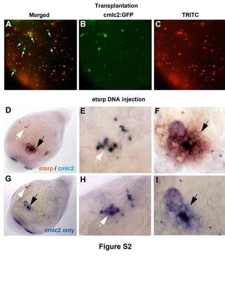
Etsrp overexpression induces ectopic cmlc2 expression in a cell non-autonomous manner. (A-C) Mosaic analysis of cmlc2:GFP expression in etsrp DNA-overexpressing embryos. Cells from cmlc2:GFP transgenic embryos injected with etsrp DNA and TRITC-dextran mixture were transplanted into uninjected cmlc2:GFP embryos at the sphere stage. Embryos were analyzed at 24 hpf for cmlc2:GFP (green) and TRITC (red) fluorescence. Merged (A), GFP (B) and TRITC (C) channels are shown. Ventral view of cells over the yolk region, anterior is upwards. Note that multiple ectopic cmlc2:GFP cells (A, arrows) do not have TRITC fluorescence. (D-F) Two-color in situ hybridization analysis of cmlc2 (blue) and etsrp (red) colocalization in etsrp DNA-injected embryos, which results in mosaic pattern of etsrp overexpression. In many cases, ectopic cmlc2-expressing cells do not overlap with etsrp expression as analyzed at the 20-somite stage. An embryo shown has both, non-overlapping (white arrow) and overlapping (black arrow) cells. Ventral view, anterior is towards the lower left. (E,F) Higher magnification views. Ectopic cmlc2/etsrp double staining in F is surrounded by brown etsrp-only stained cells. (G-I) Presence of double staining was confirmed by washing out stained embryos in ethanol, which dissolves brown iodonitrotetrazolium chloride stain but not blue nitrotetrazolium blue chloride stain. The same embryo shown as in D-F, mounting angle may be slightly different. Compare staining in E with H and F with I. |