
Fig. 4
- ID
- ZDB-FIG-111028-15
- Publication
- Palencia-Desai et al., 2011 - Vascular endothelial and endocardial progenitors differentiate as cardiomyocytes in the absence of Etsrp/Etv2 function
- Other Figures
- All Figure Page
- Back to All Figure Page
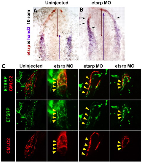
etsrp-expressing cells differentiate as cardiomyocytes in the absence of Etsrp function. (A,B) Rostrally expanded hand2 (purple) overlaps with etsrp (red) expression in Etsrp morphants (B) but not in wild-type embryos (A) at the 10-somite stage, as analyzed by two-color in situ hybridization. Red and purple arrows indicate the anterior-posterior span of etsrp and hand2 expression, respectively. Black arrows indicate the areas of overlapping expression. Ventral flat-mounted view, anterior is upwards. (C) At 30 hpf, etsrp:GFP and cmlc2:mCherry expression overlaps (yellow arrowheads) in the myocardial layer of Etsrp morphant hearts (three right columns show three different morphants) but not in control uninjected embryos (left column). Left lateral whole-mount views of fixed Tg(etsrp:GFP; cmlc2:mCherry) embryos, anterior is towards the left. Projections of only few selected slices are shown. |