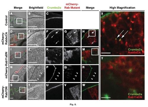
Crumbs2a localization is altered when either Rab11aDN or Rab5cCA proteins are expressed. Immunolocalization of Crumbs2a to the apical membrane in the hindbrain epithelium of (A–C) 28hpf vsx2:Gal4 control; (D–H) vsx2:Gal4;mCherry-Rab5cCA; (I–R) vsx2:Gal4/mCherry-Rab11aDN; and (S–W) vsx2:Gal4/mCherry-Rab7DN transgenic embryos. Images B,C, E–H, J–M, O–R, and U–W represent higher magnification of the boxed regions of the brightfield images in A, D, I, N, and S, respectively. Boxed regions in H and M are shown as higher magnifications in X and Y, respectively. Arrows in F–H and X indicate Crumbs2a immunostaining localized to large intracellular mCherry-Rab5cCA puncta. Hindbrain morphogenesis (asterisk in J) and Crumbs2a localization is disrupted in mCherry-Rab11aDN-expressing embryo (I–M,Y). Crumbs2a immunostaining is preserved, however, in the forebrain neuroepithelia (N–R), and in regions devoid of Rab11aDN expression as indicated by the arrowheads. S–W: Expression of mCherry-Rab7DN does not appear to affect Crumbs2a localization and expression. Scale bars in A, B, D, E, I, J, N, O, S, and T = 50 μm; scale bars in X and Y = 10 μm.
|
