
Fig. 4
- ID
- ZDB-FIG-111018-42
- Publication
- Blank et al., 2011 - A membrane-bound vertebrate globin
- Other Figures
- All Figure Page
- Back to All Figure Page
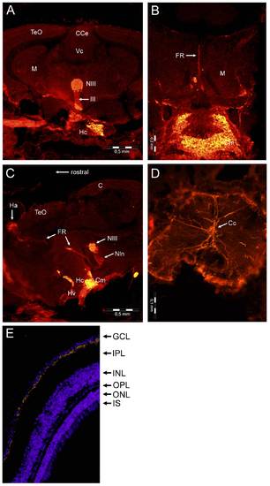
Localization of GbX in the central nervous system. Frontal section of the zebrafish brain stained with anti-GbX antibodies (A), showing the mesencephalon (M) with the tectum opticum (TeO) and the cerebellar corpus (CCe) and valvula cerebelli (Vc), two parts of the cerebellum. Staining is visible in the oculomotor nucleus (NIII) and the nervus oculomotorius (III) as well as in the caudal zone of periventricular hypothalamus (Hc). Higher magnification of stained regions of the hypothalamus (ventral) with corpus mamillare (Cm) and mesencephalon (dorsal) with fasciculus retroflexus (FR) (B). Sagittal section of the immunoreactive parts of the brain (C) with habenula (Ha), interpeduncular nucleus (Nln) and ventral zone of periventricular hypothalamus (Hv). Cross section of the spinal cord (D) showing staining in the spinal nerves of the dorsal and ventral roots. Localization of GbX in the ganglion cell layer of zebrafish retina (E). Cross section of the eye stained with the anti-GbX antibody (yellow). For staining of nuclei Hoechst 33258 was used. Ganglion cell layer (GCL), inner (IPL) and outer plexiform layer (OPL), inner (INL) and outer nuclear layer (ONL) and inner photoreceptor segment (IS). |
| Gene: | |
|---|---|
| Antibody: | |
| Fish: | |
| Anatomical Terms: | |
| Stage: | Adult |