
Fig. S7
- ID
- ZDB-FIG-111018-15
- Publication
- Patra et al., 2011 - Nephronectin regulates atrioventricular canal differentiation via Bmp4-Has2 signaling in zebrafish
- Other Figures
- All Figure Page
- Back to All Figure Page
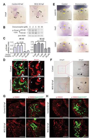
Inhibition of BMP signaling. (A) Representative images of hearts from control and MO2-injected embryos after whole-mount in situ hybridization with probes against bmp4 at 40 hpf. Note that bmp4 expression is restricted in npnt morphants at 40 hpf. Brackets indicate the AV boundary. A, atrium; V, ventricle. (B) Western blot analysis of Smad1/5/8 phosphorylation using protein extracts from 48 hpf embryos treated with dorsomorphin at 25 hpf. (C) Quantitative analysis of western blot analyses in B (mean ± s.e.m., n=4). (D) Confocal sections of whole-mount Alcam-stained (red) control and 10 μM dorsomorphin-treated embryos from transgenic zebrafish Tg(kdrl:EGFP)s843 at 52 hpf. The number of differentiated endocardial cells (cuboidal in shape, Alcam-positive and EGFP-positive) remained unchanged in dorsomorphin-treated animals. Arrowheads indicate the AV boundary. SAV, superior AV; IAV, inferior AV. (E) Bright-field images of 52 hpf control and 10 μM dorsomorphin-treated embryos after whole-mount in situ hybridization with probes against notch1b, has2 and cspg2. Expression of these genes remained unaffected in dorsomorphin-treated embryos. (F) Sections (2 μm) of the heart from control and MO2-injected embryos after whole-mount bmp4 in situ hybridization at 52 hpf. EC, endocardium; MC, myocardium. (G,H) Confocal sections of whole-mount p-Smad1/5/8-stained (red) control and MO2-injected embryos from transgenic Tg(kdrl:EGFP)s843 zebrafish at 52 hpf. Note that p-Smad1/5/8 staining in control hearts is weaker in the AV endocardium (green) compared with the myocardium. By contrast, in npnt morphants the p-Smad1/5/8 staining is of similar intensity in the expanded AV endocardium and myocardium. Scale bars: 50 μm in A,D,E,G,H; 20 μm in F. |