Fig. 10
- ID
- ZDB-FIG-111011-12
- Publication
- Stevens et al., 2011 - Plasticity of photoreceptor-generating retinal progenitors revealed by prolonged retinoic acid exposure
- Other Figures
- All Figure Page
- Back to All Figure Page
|
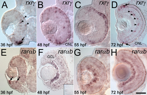
Specific retinoic acid receptors are expressed in the embryonic zebrafish retina. Wild-type, untreated embryos were fixed at 36 hpf (A, E), 48 hpf (B, F), 55 hpf (C, G), or 72 hpf (D, H), and were hybridized as 3 μm cryosections with probes corresponding to RXRγ (A-D) or RARαb (E-H); dorsal is up in all panels. (A to D) In younger embryos, RXRγ is expressed at the outer edge of ventral retina (arrow in A), and in scattered cells in inner and outer retina (arrowheads in A). In older embryos this expression becomes restricted to the emerging outer nuclear layer (ONL, B and C), and then to the most peripheral cells of the outer nuclear layer (black arrows in D) and periodically distributed cells of the inner nuclear layer (arrowheads in D). E.-H. In younger embryos, RARαb is expressed at the inner edge of ventral retina (arrows in E), and in older embryos shows widespread expression throughout the retina, with strongest hybridization signals in the ganglion cell layer (GCL) (F-H). The inset in F shows a representative control experiment using "sense" strand RARαb cRNA. Bar = 50 μm. |

