
FIGURE
Fig. 6
- ID
- ZDB-FIG-110812-22
- Publication
- Liu et al., 2011 - Cell adhesion molecule cadherin-6 function in zebrafish cranial and lateral line ganglia development
- Other Figures
- All Figure Page
- Back to All Figure Page
Fig. 6
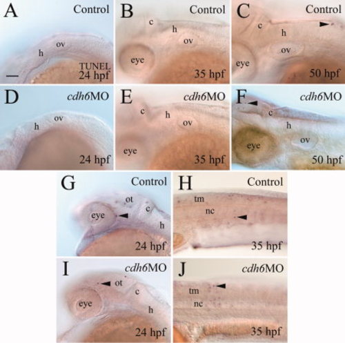
Apoptosis analysis using TUNEL (terminal deoxynucleotidyl transferase–mediated deoxyuridinetriphosphate nick end-labeling) staining. All panels show lateral views of whole-mount embryos (anterior to the left and dorsal up) processed for TUNEL staining. The mid- and hindbrain region focusing on the otic vesicle (A–G,I), while panels H and J are from the body trunk region (H,J). Arrowheads point to some TUNEL-positive cells. nc, notochord; tm, trunk muscles. Other abbreviations are the same as in Figure 1. Scale bar = 50 μm for all panels. |
Expression Data
Expression Detail
Antibody Labeling
Phenotype Data
| Fish: | |
|---|---|
| Knockdown Reagent: | |
| Observed In: | |
| Stage Range: | Prim-5 to Long-pec |
Phenotype Detail
Acknowledgments
This image is the copyrighted work of the attributed author or publisher, and
ZFIN has permission only to display this image to its users.
Additional permissions should be obtained from the applicable author or publisher of the image.
Full text @ Dev. Dyn.