
Fig. 3
- ID
- ZDB-FIG-110804-61
- Publication
- Saito et al., 2011 - Isolation and cytogenetic characterization of zebrafish meiotic prophase I mutants
- Other Figures
- All Figure Page
- Back to All Figure Page
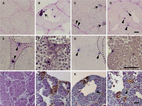
Apoptotic cells in the its, isa, and imo mutant testes. A–H: Immunohistochemical staining of cleaved Caspase-3 in wild-type (A), its (B), isa (C, E, and F), and imo (D,G,H) testes. The testes sections were stained with anti-cleaved Caspase-3 antibodies, and counterstained with periodic acid Schiff. Immunological reactivity was observed in germ cells in its (B), both germ cells and Leydig cells in isa (C), and Sertoli cells in imo (D). High magnification view of hematoxylin counterstaining images (F,H) are same position as seen in cleaved Caspase-3 positive cells in isa (E) and in imo (G). Dotted line indicates the position of the basement membrane. Arrows indicate apoptotic germ cells (B and C). Arrowheads indicate apoptotic Leydig cells (C,E) and Sertoli cells (D,G), respectively. I–L: TUNEL (terminal deoxynucleotidyl transferase–mediated deoxyuridinetriphosphate nick end-labeling) staining of wild-type (I), its (J), isa (K), and imo (L) testes. Wild-type testis shows a very low incidence of apoptosis. In contrast, the high incidence of apoptotic TUNEL staining (brown) was observed in germ cells of all three mutant testes. In addition, apoptotic TUNEL staining was also observed in Leydig cells (K) and Sertoli cells (L). Arrowheads indicate apoptotic Leydig cells (K) and Sertoli cells (L), respectively. Scale bars = 25 μm. |
| Fish: | |
|---|---|
| Observed In: | |
| Stage: | Adult |