
Fig. s3
- ID
- ZDB-FIG-110622-54
- Publication
- Takahashi et al., 2011 - An Enzymatic Mechanism for Generating the Precursor of Endogenous 13-cis Retinoic Acid in the Brain
- Other Figures
- All Figure Page
- Back to All Figure Page
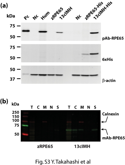
Specificity of the anti-RPE65 antibodies to recombinant zebrafish 13cIMH. (a) The expression plasmids were transfected into 293A cells and cultured for 48 hours. The cells were harvested, equal amounts (20 μg) of total cellular protein were resolved by SDS-PAGE, and the protein expression was confirmed by Western blot analysis with a polyclonal anti-human RPE65 [4], mouse monoclonal anti-His-tag, and goat polyclonal anti-β-actin antibodies at same time. Pc, positive control (bovine RPE microsomal protein); Nc negative control (cells expressing RFP); Hum, human RPE65; zRPE65, zebrafish RPE65 without 6xHis-tag; 13cIMH, zebrafish 13cIMH without 6xHis-tag; zRPE65-His, zebrafish RPE65 with 6xHis-tag; 13cIMH-His, zebrafish 13cIMH with 6xHis-tag. Subcellular fractionation was performed as explained in Materials and Methods. The polyclonal antibody recognized zebrafish 13cIMH but not zebrafish RPE65. (b) The cells expressing 13cIMH were fractionated. The same amounts of protein from each fraction (5 μg) were blotted with a monoclonal anti-human RPE65 antibody (Millipore, Billerica, MA) and a rabbit polyclonal antibody to calnexin (ER membrane marker, Abcam, Cambridge, MA). The monoclonal antibody recognized zebrafish 13cIMH but not zebrafish RPE65 (zRPE65). T; total cell lysates, C; cytosolic, M; membrane, N; nuclear fractions and S; detergent-insoluble fraction including cytoskeleton and inclusion body. |