
Fig. 3
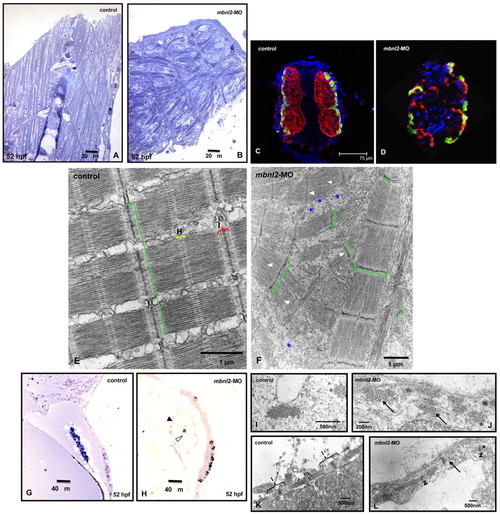
Histological and molecular analyses of skeletal and cardiac muscle of mbnl2 morphants. (A,B) In longitudinal sections through the muscles of the tail of zebrafish embryos at 40× magnification, the controls (A) show a normal somite chevron shape and fibre alignment, whereas the mbnl2 morphants (B) show an abnormal somite shape, and misaligned muscle fibres, which appear scattered in the somitic compartment. (C,D) Immunofluorescence staining with S58 and MF20 antibodies in transverse sections of 52-hpf zebrafish embryos shows slow (S58 positive; yellowish green) and fast (S58 negative, MF20 positive; red) fibre distribution in somitic muscles of control embryos (C) compared with mbnl2 morphants (D); DAPI was used as a blue nuclear counterstain. (E) Electron micrographs of skeletal muscle from 52-hpf control zebrafish show that the myofibrils are aligned, their Z-bands are regularly stacked (green square bracket), and H-zones (H) and I-bands (I) are clearly identifiable. (F) mbnl2 morphants show misaligned myofibrils, Z-bands with irregular width, disorganized sarcoplasmic reticulum (*) and short sarcomeres resulting in loss of H-zones and I-bands (arrowheads). (G) In normal zebrafish hearts (20× magnification), the blood cells fill in the atrial cavity (a). (H) By contrast, the heart in mbnl2 morphants is dilated and empty; black arrowhead points to the myocardium and white arrowhead to the endocardium. (I–L) Electron micrographs of sections through the hearts of zebrafish embryos. Tightly organized myofibril bundles can be seen in transverse sections of wild-type controls (I), whereas dispersed myofibrils (arrows) are apparent in the mbnl2 morphant embryos (J). (K) Longitudinal sections showing normal sarcomeric organization of the Z-line (z), H-zone (h), I-band (I) and M-line (m) in controls. (L) The myofibrils are less organized and sometimes disrupted (arrow) in the morphants, condensation of the Z-bands in the sarcomere can be seen (*), and H-zone and I-band are not recognizable in severe cases. |
| Antibodies: | |
|---|---|
| Fish: | |
| Knockdown Reagent: | |
| Anatomical Terms: | |
| Stage: | Long-pec |