
Fig. S1
- ID
- ZDB-FIG-110608-24
- Publication
- Tu et al., 2011 - Fate restriction in the growing and regenerating zebrafish fin
- Other Figures
- All Figure Page
- Back to All Figure Page
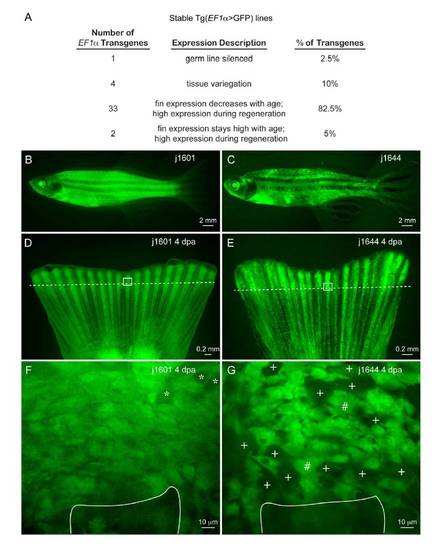
Stable transgenic EF1α>GFP lines express GFP ubiquitously in the caudal fin. (A) Summary of 40 EF1α>GFP stable lines generated using Tol2 EF1α>GFP lineage marking transposon. Almost 90% of the stable transgenic lines have uniform expression in the adult caudal fin, while 10% of the lines exhibit variegating GFP expression. Examples of GFP expression in adults in a uniform expressing line (B, D, F) and in a line that shows tissue variegation (C, E, G). Following amputation, GFP expression is typically up-regulated in all cells of the regenerate in both uniform lines (D) and variegating lines (E), similar to a previously reported stable EF1α>GFP transgenic line (Thummel et al., 2006). (F,G) Representative confocal, sagittal Z-sections through 4 dpa proximal regenerates in the uniform expression line and the variegating expression line corresponding to areas outlined by white boxes in whole mounts (D, E) shown above. Asterisks indicate vascular lumen and white lines outline the positions of fin rays in each stump, with top of line indicating the amputation plane. In the uniform expression line, every cell expresses GFP, which tends to rule out the possibility that clonal analysis missed a role for transdifferentiation due to transdifferentiation-dependent EF1α>GFP transgene silencing. In the variegating fin regenerate (H), we observed the majority of cells showed strong GFP expression (#) while a minority of cells showed dimmer GFP expression (+), at levels that would likely be detected in clonal analysis. EF1α>GFP stable transgenic lines are available from the Zebrafish International Resource Center. |
Reprinted from Developmental Cell, 20(5), Tu, S., and Johnson, S.L., Fate restriction in the growing and regenerating zebrafish fin, 725-732, Copyright (2011) with permission from Elsevier. Full text @ Dev. Cell