
FIGURE
Fig. 4
- ID
- ZDB-FIG-110608-23
- Publication
- Tu et al., 2011 - Fate restriction in the growing and regenerating zebrafish fin
- Other Figures
- All Figure Page
- Back to All Figure Page
Fig. 4
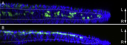
Confocal Microscopy Shows Spatial Restriction of Dermal Fibroblasts and Osteoblast in the Regeneration Blastema (A) Dermal fibroblast clone in 4 days postamputation (dpa) regeneration blastema. (B) Osteoblast clone in 4 dpa regeneration blastema. Dotted line outlines the basement membrane. Blue shows DAPI stain and green illustrates GFP (immunohistochemistry). L and R denote left and right sides of fish, respectively. |
Expression Data
Expression Detail
Antibody Labeling
Phenotype Data
Phenotype Detail
Acknowledgments
This image is the copyrighted work of the attributed author or publisher, and
ZFIN has permission only to display this image to its users.
Additional permissions should be obtained from the applicable author or publisher of the image.
Reprinted from Developmental Cell, 20(5), Tu, S., and Johnson, S.L., Fate restriction in the growing and regenerating zebrafish fin, 725-732, Copyright (2011) with permission from Elsevier. Full text @ Dev. Cell