|
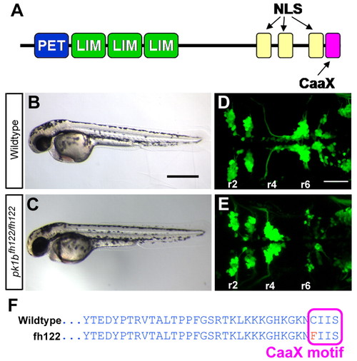
prickle1b is disrupted in zebrafish fh122 mutants. (A) The domain structure of Pk1b. NLS, nuclear localization signal. (B,C) Lateral brightfield views of 48 hpf wild-type (WT) (A) and pk1bfh122/fh122 (B) embryos. pk1bfh122/fh122 embryos are morphologically normal. Anterior is to the left. (D,E) Maximum projection dorsal views of cranial branchiomotor neurons (CBMNs) in 48 hpf Tg(islet1:GFP) embryos. Rhombomeres are indicated (r2-r6). In WT embryos (C), facial branchiomotor neuron (FBMN) cell bodies migrate from r4 to r6. In fh122 mutants (D), FBMNs fail to undergo caudal tangential migration and cell bodies cluster in r4. (F) Amino acid sequence of Pk1b in WT and pk1bfh122/fh122 fish. The fh122 mutation results in a C-to-F transition in the Pk1b farnesylation (CaaX) motif. Scale bars: 500 μm in B; 50 μm in D.
|