
Fig. 3
- ID
- ZDB-FIG-110526-5
- Publication
- Fogelgren et al., 2011 - The Exocyst Protein Sec10 Interacts with Polycystin-2 and Knockdown Causes PKD-Phenotypes
- Other Figures
- All Figure Page
- Back to All Figure Page
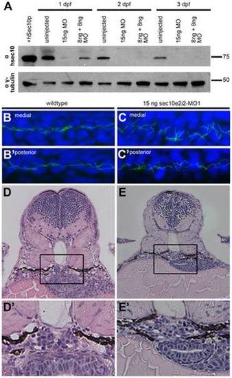
sec10MO embryos show abnormal pronephric development. (A) Immunoblots showing the 1-3 dpf time course of zfSec10 knockdown in sec10MO embryos. sec10MO injections can lead to abnormal-appearing embryos (Table 1), but in this blot, 3 dpf sec10MO embryo lysates were isolated only from embryos without any obvious morphological defects. This demonstrates that these embryos still have strong knockdown of the zfSec10 protein. Lysates from abnormal-appearing embryos showed similar levels of zfSec10 knockdown (data not shown). 5 embryos loaded per lane. Positive control for Sec10 was from human Sec10 (hSec10) overexpressing MDCK cell lysates. Blot was probed with antibodies against hSec10, and gamma-tubulin as a loading control. (B-C′) Immunofluorescence with antibody against acetylated-tubulin (green), and the nuclear Hoechst stain (blue). Flattened Z-series from confocal imaging of medial (B,C) and posterior kidney (B′,C′), 24 hours post fertilization (hpf), lateral view, 80x magnification. Pronephric cilia length is similar between uninjected embryos (B/B2) and 15ng sec10MO embryos (C/C′); however, cilia within the medial pronephros are disordered (compare B and C). (D-E′) JB-4 resin section (with enlarged inset) of glomerular region, stained with Hematoxylin and Eosin, 3 dpf, transverse 4 μm section, 40x magnification. Wild-type embryos show an organized U-shaped glomerulus (D/D′), while 15ng sec10MO embryos show disorganization (E/E′). |
| Fish: | |
|---|---|
| Knockdown Reagents: | |
| Observed In: | |
| Stage Range: | Prim-5 to Protruding-mouth |