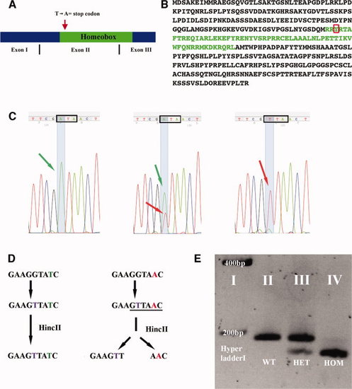
evx1 mutation probably causes a complete loss of function. A: Schematic of evx1 cDNA indicating exon boundaries. Red arrow shows approximate position of point mutation that converts a Thymine to Adenine and, hence, the Tyrosine (red box in B) into a stop codon. B: Amino acid sequence of Evx1, homeobox in green. C:evx1 sequencing results (reverse complement) for wild-type (WT; left), heterozygous (middle) and homozygous mutant (right) zebrafish. Green arrows indicate Thymine (reverse complement = A; WT allele); Red arrows indicate Adenine (reverse complement = T; mutant allele). Black box outlines the codon that produces Tyrosine in WTs and an ochre stop codon in mutants. Note the change in sequence (SYR [AGT TAT CGA] instead of RYR [AGG TAT CGA]) deliberately introduced by mismatch primers during polymerase chain reaction (PCR; see the Experimental Procedures section). D: Schematic of evx1 mutant genotyping. WT allele left, mutant right. Thymine (purple) is substituted for Guanine by means of PCR. This creates a HincII site (underlined) in mutant DNA. Subsequent enzymatic digestion results in two fragments (158 bp and 28 bp) in mutants while leaving WT DNA uncut (186 bp). E: Genotyping results visualized by gel electrophoresis. The 28-bp fragment generated by HincII digest of mutant DNA is not visible.
|
