
FIGURE
Fig. 4
- ID
- ZDB-FIG-110517-3
- Publication
- Matsuda et al., 2011 - Detection of vitellogenin incorporation into zebrafish oocytes by FITC fluorescence
- Other Figures
- All Figure Page
- Back to All Figure Page
Fig. 4
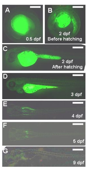
Development of zebrafish embryos that incorporated FITC-labeled Vtg, heterogeneously prepared from the bubble-eye goldfish, in the yolk during oocyte growth. Embryonic stage is given as days post fertilization (dpf). Scale bar = 200 μm. |
Expression Data
Expression Detail
Antibody Labeling
Phenotype Data
Phenotype Detail
Acknowledgments
This image is the copyrighted work of the attributed author or publisher, and
ZFIN has permission only to display this image to its users.
Additional permissions should be obtained from the applicable author or publisher of the image.
Full text @ Reprod. Biol. Endocrinol.