
Fig. 2
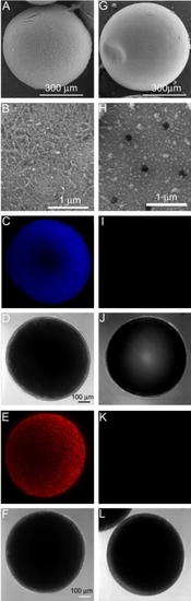
Representative surface images of follicle-enclosed stage IV oocytes (A–F) compared to denuded stage IV oocytes (G–L), photographed using a scanning electronic microscope (SEM) or a confocal scanning microscope. (A), (B), (G) and (H) are recorded under the SEM; (B) and (H) show the magnified surface of the same follicle-enclosed oocyte in picture A or the denuded oocyte in picture G. Images of (C–F) and (I–L) were photographed under a confocal microscope. (C), (E), (I) and (K) are projected confocal images of follicle-enclosed oocytes (C and E) compared to denuded oocytes (I and K) stained with 4′,6-diamidino-2-phenylindol (DAPI, C and I) or propidium iodide (PI, E and K). (D), (F), (J) and (L) were taken using differential interference contrast (DIC), corresponding to the same oocytes in the pictures directly above (C), (E), (I) and (K), respectively. |
Reprinted from Molecular and Cellular Endocrinology, 337(1-2), Hanna, R.N., and Zhu, Y., Controls of meiotic signaling by membrane or nuclear progestin receptor in zebrafish follicle-enclosed oocytes, 80-8, Copyright (2011) with permission from Elsevier. Full text @ Mol. Cell. Endocrinol.