
|
Fig. 2
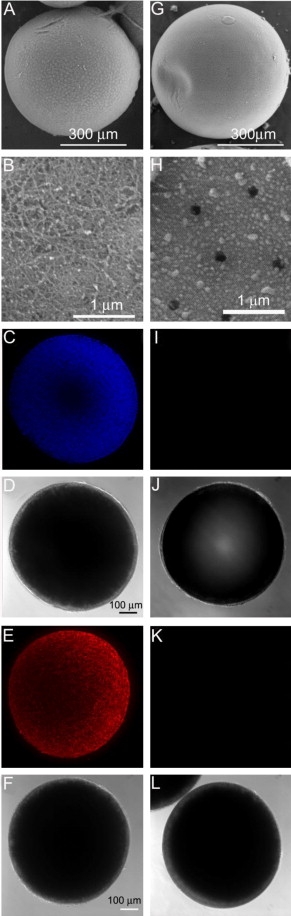
Representative surface images of follicle-enclosed stage IV oocytes (A–F) compared to denuded stage IV oocytes (G–L), photographed using a scanning electronic microscope (SEM) or a confocal scanning microscope. (A), (B), (G) and (H) are recorded under the SEM; (B) and (H) show the magnified surface of the same follicle-enclosed oocyte in picture A or the denuded oocyte in picture G. Images of (C–F) and (I–L) were photographed under a confocal microscope. (C), (E), (I) and (K) are projected confocal images of follicle-enclosed oocytes (C and E) compared to denuded oocytes (I and K) stained with 4′,6-diamidino-2-phenylindol (DAPI, C and I) or propidium iodide (PI, E and K). (D), (F), (J) and (L) were taken using differential interference contrast (DIC), corresponding to the same oocytes in the pictures directly above (C), (E), (I) and (K), respectively.
Reprinted from Molecular and Cellular Endocrinology, 337(1-2), Hanna, R.N., and Zhu, Y., Controls of meiotic signaling by membrane or nuclear progestin receptor in zebrafish follicle-enclosed oocytes, 80-8, Copyright (2011) with permission from Elsevier. Full text @ Mol. Cell. Endocrinol.