
FIGURE
Fig. S3
Fig. S3
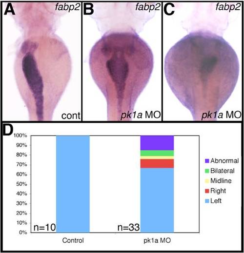
Abnormal intestine localization in pk1a morphants. Whole-mount in situ hybridization of fatty acid binding protein 2 (fabp2), an intestinal marker, in 3 dpf control (A) and pk1a morphants demonstrates abnormal intestine location, including “bilateral” (B), and “right” (C). (D) Graph depicting the scoring of pk1a morphants demonstrates a significant difference in the number of abnormally localized intestines (0% vs. 33%, p<0.0001 by chi-square test). Note that the fabp2 probe also faintly stains the liver. |
Expression Data
Expression Detail
Antibody Labeling
Phenotype Data
| Fish: | |
|---|---|
| Knockdown Reagent: | |
| Observed In: | |
| Stage: | Protruding-mouth |
Phenotype Detail
Acknowledgments
This image is the copyrighted work of the attributed author or publisher, and
ZFIN has permission only to display this image to its users.
Additional permissions should be obtained from the applicable author or publisher of the image.
Reprinted from Developmental Biology, 351(2), Cui, S., Capecci, L.M., and Matthews, R.P., Disruption of planar cell polarity activity leads to developmental biliary defects, 229-241, Copyright (2011) with permission from Elsevier. Full text @ Dev. Biol.