
Fig. 4
- ID
- ZDB-FIG-110321-6
- Publication
- Wibowo et al., 2011 - Compartmentalized Notch signaling sustains epithelial mirror symmetry
- Other Figures
- All Figure Page
- Back to All Figure Page
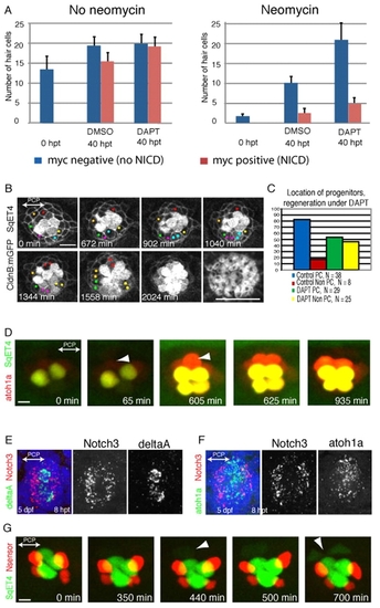
UHCP develop in polar compartments. (A) Quantification of the average number of hair cells in samples that were treated with neomycin (right) or that were not treated (left), and later exposed to DMSO or DAPT, and that expressed (red) or did not express (blue) a constitutively active form of Notch. It shows that DAPT induces the overproduction of hair cells, and this effect is suppressed by Notch activity. Results are mean±s.e.m. (B) A 34-hour time series of a regenerating Tg[Cldnb:mGFP; SqET4] neuromast treated with DAPT. UHCPs were identified retrospectively by playing the time series backwards, and labeled with colored dots. UHCPs develop in excess, producing pairs of hair cells ectopically. Actin staining shows the resulting defective planar polarization (last panel). (C) A graph showing the position of UHCPs relative to the polar compartments in control and DAPT-treated neuromasts during regeneration. More than 80% of UHCPs develop within the polar compartments in controls, compared with just over 50% in DAPT-treated samples (n=25). Results are mean±s.e.m. (D) Time series of a Tg[Atoh1a:TdTomato; SqET4] double-transgenic neuromast revealing GFP-positive UHCPs and hair cells (green), and Atoh1a-expressing cells (red). It shows the temporal gene-expression hierarchy. Arrowheads indicate UHCPs. (E,F) Fluorescent whole-mount in situ hybridizations of regenerating neuromasts, revealing Notch3 (red) and DeltaA (green) (E), and Notch3 (red) and Atoh1a (green) (F). Cell nuclei are in blue. Notch3 is never expressed by the DeltaA(+) or Atoh1a(+) cells, and was absent from the polar compartments. (G) A 700-minute live imaging of a double-transgenic SqET4 (green) neuromast expressing a red-fluorescent Notch sensor (red). Notch signaling occurs outside the polar compartments. Arrowheads indicate UHCPs. |